Protein Story
Tus Replication-Terminator Protein
|
|
Protein Story Tus Replication-Terminator Protein |
In the Escherichia coli chromosome, bidirectional DNA replication is initiated from a single orgin. Two replication forks, driven by the replication machinery, travel in opposite directions around the circular chromosome and terminate in a region opposite the origin. Two components dictate the arrest of the progression of replication. One is a set of six DNA sequences, designated as Ter (Fig.1), each containing a element about 20 base pair long. The other is a trans-acting protein encoded by tus gene.The tus protein (Tus, Mt=36K) specifically binds to each Ter site as a monomer, forming a protein-DNA complex. This complex halts the passage of the replication fork in only one direction and permits passage by the other replication fork. The Ter sites are oriented so as to restrain DNA replication proceeding in the terminus-to- origin direction. Thus, this polar fork-blocking mechanism ensures that the terminus region works as a replication-fork trap.
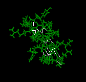
Now, let's go further to find out what is Tus protein looks like. The sequence and the secondary structure of Tus are shown in Fig.2, and the ribbon drawing of the Tus-DNA complex is shown in Fig.3, Fig.4. The structure is divided into two domain (amino and carboxy). This overall structure has three distinct regions: two alpha-helical regions and central beta structures, which jointly form a central large cleft. The Ter DNA is accommodated into the positively charged central cleft, as the two protruding alpha-hilical regions and the core beta structures embrace 13bp of the DNA duplex. There are so many stories about each subdomains of this structure, from alpha I to alpha VII, from beta A to beta O and from L1 loop to L4 loop.....wow!! But we will choose the most important part, also the most interesting part, for searching further:
Three amphipathic alpha-helices (alpha I, II and III) form an antiparallel helix bundle aligned roughly in parallel with the DNA. Two alpha IV and V helices, which are connected by a turn across a DNA backbone, clamp the phosphate backbones from both brooves (Fig.4).
This is the most interesting part of the whole structure. The two domains are connected mainly through twisted, antiparallel beta-strands (beta F-G and beta H-I). These interdomain beta-strands, which are portions of two beta-sheets of the two domains, contribute to the extensive contact with the bases of the major groove and the sugar-phosphate backbones of the Ter DNA.
The L4 loop (residues 283-296), which joint with interdomain beta-strands, is involved in the base recogmition. And the L3 loop (residues 196-204) which between the two alpha-helices contacts the minor groove of the DNA.
Tus has a proline repeat (Fig.5) which can be found in many DNA-binding protein. This kind of repeat may involve protein/protein interactions between the Tus-DNA complex and subunits of replicase. At the 178 amino acid C-terminal segment is punctuated by several helix-destabilizing glycine and proline residues.
Ok, it's no time now. The story of the lovely Mr.Tus must be ended. As we go through the above stuff, we can feel that we still not know it well. About it's family about it's history.....but it doesn't matter, we still have much time to be with it ......