Find a protein structure from PDB.
Show it on your homepage with three GIF pictures.
Give some references and bckground of this protein.


     Compound: Zine finger
ID: 3znf
Method: NMR
    
Reference:
High-resolution three-dimensional structure of a single zinc finger from a human enhancer binding protein in solution. J. G. Omichinski, G. M. Clore, E. Appella, K. Sakaguchi & A. M.Gronenborn Biochemistry 29: 9324-34 (1990)
Detail Information

         
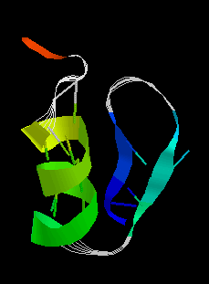
The basic structure: 1 helix, 2 strands, 3 turns, and 15 H-bonds.
   
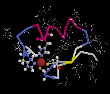
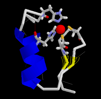
In this picture, the 4 residues in "ball&stick" interact with the ligand, which is in red.


The blue ribbon is helix. The yellow strands are sheet. The red ball is ligand. And the 4 residues are His21, His 27, Cys 5, and Cys 8, which interact with the ligand.