|
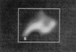
2-dimensional projection map of one 50S ribosomal subunit at a resolution of 30 Angstrom. The protrusion (S) can be more readily recognizable from the map. |
 |
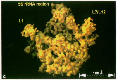 |
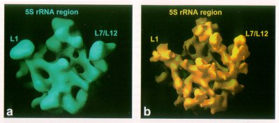
| Comparison of the 20 Angstrom resolution EM reconstruction with both 20 Angstrom (a) and 12 Angstrom (b) resolution X-ray maps of the HM 50S subunit |
 |
Selected views of the 70S ribosome of E. coli as obtained by cryo-electron microscopy of single ribosomes and reconstruction using 4300 projections (Frank et al., Nature 376 (1995) 441-444). Yellow: 30S subunit, blue: 50S subunit. |
 |
Left panel: reconstruction cut open, shown with interpretative elements (mRNA, A- and P-site tRNAs and two possible pathways of the polypeptide chain). Right panel: uncut reconstruction, shown with the same orientations as in the panel on the left. Upper left: mRNA is thought to enter through the channel, present message in the region of the cleft, and exit between head and platform of the 30S subunit. Middle left and bottom left: tRNAs shown in two extremes of a range of positions, related by rotation of the anticodon ends around the mRNA "axis". Landmarks of the 30S subunit: h, head; p, platform; ch, channel presumed to be the conduit for mRNA; sp, spur. Landmarks of the 50S subunit: CP, central protuberance; St, L7/L12 stalk; L1, L1 protein; IC, interface canyon; T, tunnel presumed to be the conduit for the nascent polypeptide chain; T1 and T2, lower tunnel segments, leading to alternative exit sites E1 and E2, respectively. (from Frank et al., Biochem. Cell Biol. 73 (1995) 757-765.) |