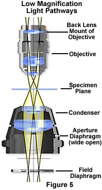
The modified optical pathway for transmitted light
in a low power microscope system is illustrated in Figure 5. The field
diaphragm is now positioned in the correct plane to act as an aperture
stop to control the numerical aperture and the size and shape of the light
cone for the illuminating and image-forming light rays passing through
the condenser. The specimen plane is fully illuminated with equiangular
cones (as in Kohler illumination), however the filament is now imaged at
back lens mount of the objective instead of the back focal plane.