The resolution of a microscope objective
is defined as the smallest distance between two points on a specimen that
can still be distinguished as two separate entities. Resolution is a somewhat
subjective value in microscopy because at high
magnification, an image may appear
unsharp but still be resolved to the maximum ability of the objective.
Numerical aperture determines the resolving power of an objective, but
the total resolution of a microscope system is also dependent upon the
numerical aperture of the substage condenser. The higher the numerical
aperture of the total system, the better the resolution.
The numerical aperture (N.A.) is basically a value that
describes the quality of a lens. It is derived from the size of the lens,
its working distance and the index of refraction. All quality objective
lens will state the numerical aperture on the side of the barrel. A good
rule of thumb is that the effective magnification of an objective is its
numerical aperture times 1000. So a 40 x objective that has a N.A. of 0.65
has an effective magnification of 650 times. If you magnify beyond this
you will only get empty magnification. You can calculate the theoretical
resolution of any optical system using Abbe's equation. To calculate the
resolution of the objective above multiple the wavelength of green light(0.5
micrometers) times the constant .61 divided by the N.A. The result will
be 0.47 micrometers. In another example you can calculate the resolution
of a pair of 8 x 20 binoculars. The number 8 is the magnification and the
number 20 is the diameter of the objective lens. Assume you were looking
at a specimen 100 ft away the alpha would be 0.0188 degrees. Plugging in
abbe's equation the result for red light (650 nm) is 1.2 mm. Remember,
this is a theoretical value with is the best possible resolution possible.
The practical resolution will always be less due to optical aberratio.
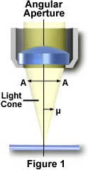
The angle m is one-half the angular aperture (A) and is related to the numerical aperture through the following equation:
Numerical Aperture (NA) = n(sin m)
where n is the refractive
index of the imaging medium between the front lens of the objective
and the specimen cover glass, a value that ranges from 1.00 for air
to 1.51 for specialized immersion oils. From this equation it is
obvious that when the imaging medium is air (with a refractive index,
n = 1.0), then the numerical aperture is dependent only upon the angle
m whose maximum value is 90°. The sin of the angle m, therefore,
has a maximum value of 1.0 (sin(90°) = 1), which is the theoretical
maximum numerical aperture of a lens operating with air as the imaging
medium (using "dry" microscope objectives).
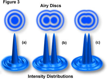
An important concept to understand
in image formation is the nature of diffracted light rays intercepted by
the objective. Only in cases where the higher (1st, 2nd, 3rd, etc.) orders
of diffracted rays are captured, can interference work to recreate the
image in the intermediate image plane of the objective. When only the zeroth
order rays are captured, it is virtually impossible to reconstitute a recognizable
image of the specimen. When 1st order light rays are added to the zeroth
order rays, the image becomes more coherent, but it is still lacking in
sufficient detail. It is only when higher order rays are recombined, that
the image will represent the true architecture of the specimen. This is
the basis for the necessity of large numerical apertures (and subsequent
smaller Airy disks) to achieve high-resolution images with an optical microscope.