![]() due on
5/28
due on
5/28
![]() 1. David Liu,
same guy from last homework, also isolated a cDNA clone from snake
venom library. The nucleotide sequence of this cDNA clone is shown
here:
1. David Liu,
same guy from last homework, also isolated a cDNA clone from snake
venom library. The nucleotide sequence of this cDNA clone is shown
here:
AAAACCATCAAATATGTTATGCTGGAATGCAACGAACTGATCCCGCTGTTC
TACGAAACCTGCCCGGCTGGTGAAAACATCTGCTACGAAATGTTCATGGTT
GCTACCCCGAAAGTTCCGTGCGAACGTGGTTGCATCGACGTTTGCCCGGAA
TCTTCTCTGATCGTTAAATACGTTTGCTGCAACACCGACCGTTGCCAGTAAT
CCAGCGCCTGATCTCTCGAAATAAAAGCCGCATTG
(1) Please help him to find its corresponding polypeptide sequence (DNA -> Protein translation).
(2) Please help him to calculate pI/Mw of this polypeptide, perform the trypsin cutting , analyze the cutting pattern and report the fragments with molecular weight over 500 Dalton.
(3) Please help him to identify this toxin. Is it a new toxin?
(4) Please help him to use Prosite scanning tool to find out possible functions or pattern of this polypeptide.
(5) David would like to see its structure. Could you help him to find structure of this toxin or make a model if it is a new protein? Show structure on your homwpage ( 3 different views).
Ans:
(1) We used the Expasy and could translate the DNA sequence to protein sequence. 6 frames were gotten here.
The 5'-3' frame 1 has the longest openreeading frame and shown below:
K T I K Y V Met L E C N E L I P L F Y E T C P A G E N I C Y E Met F Met V A T P K V P C E R G C I D V C P E S S L I V K Y V C C N T D R C Q Stop S S A Stop S L E I K A A L
(2) Theoretical pI/Mw: 4.29 / 6917.13.
The peptide masses which are over 500 Da after treated with trypsin are listed below:
mass position peptide sequence
---------- --------- ----------------
3699.66 1- 32 MLECNELIPLFYETCPAGENICYEMFMVATPK
1462.73 38- 51 GCIDVCPESSLIVK
973.39 52- 59 YVCCNTDR
603.29 33- 37 VPCER
(3) A 98% homologous protein is found...
(4) Use the prosite scaning tool can get two possible functions in this protein. One is protein kinase C phosphorylation site and the other is snake toxin signature.
(5) Three views in putative structure:
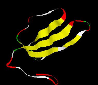
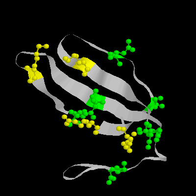
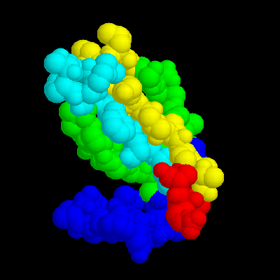
In this putative structure shown here has four beta-sheets (Fig.1 in yellow), and charged residues labeled in Fig.2. Fig.3 in colored in grouply. You can link the PDB file here.
![]()