due on 4/29
![]() 1.
Find the structure of trp repressor
complexed with its operator from PDB. List the following information
: (1) ID number, (2) Authors, (3)
Resolutions, (4) How many residues in one monomer,
(5) How many cofactors in whole complex. Show the complex molecule on your
homepages. Show different color for different chain in cartoon form.
Indicate all the cofactors in spacefill form and blue color. Show the
script that makes the final picture.
1.
Find the structure of trp repressor
complexed with its operator from PDB. List the following information
: (1) ID number, (2) Authors, (3)
Resolutions, (4) How many residues in one monomer,
(5) How many cofactors in whole complex. Show the complex molecule on your
homepages. Show different color for different chain in cartoon form.
Indicate all the cofactors in spacefill form and blue color. Show the
script that makes the final picture.
Ans:
(1) ID number: 1tro.
(2) Authors: Z.OTWINOWSKI,R.G.ZHANG,P.B.SIGLER.
(3) Resolutions: 1.9 ANGSTROMS .
(4) number of residues per monomer: 108.
(5) number of cofactors in whole complex: 4 .
ps: The original download is linked from PDB www site here.
The requested image and script are shown, too.

![]() 2. Transcription of the ant gene during lytic
growth of bacteriophage P22 is regulated by the cooperative binding
of two Arc repressor dimers to a 21-base-pair operator site. Please
find the co-crystal
structure of this Arc tetramer-operator
complex from PDB.
2. Transcription of the ant gene during lytic
growth of bacteriophage P22 is regulated by the cooperative binding
of two Arc repressor dimers to a 21-base-pair operator site. Please
find the co-crystal
structure of this Arc tetramer-operator
complex from PDB.
(a) Make an animation (by Gifbuilder) of whole complex.
(b) Show the DNA sequences of the Arc operator.
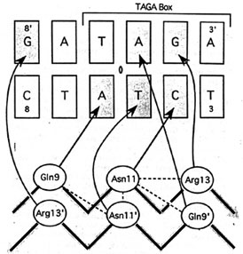
(c) The picture below is the hydrogen bonds between beta-sheet side chains and DNA bases. Show three side chains (gln9, asn11, arg13) from one monomer and corresponding DNA bases of them on your home page. Color three side chain with different color. Measure the distance between possible hydrogen bonds.
Ans:
(1) The animation of whole complex...
.
(2)DNA sequence:
SEQRES 1 E 5' TATAGTAGAGTGCTTCTATCAT 3'
SEQRES 1 F 5' AATGATAGAAGCACTCTACTAT 3'
(3) The figures were shown. Three h-bonds shown together, or G4E-Arg13D, T17E-Asn 11D, A7F-Gln9D shown individually can make clear views to measure the h-bonds.
![]()