Pedro M. Coutinho and Peter J. Reilly
Department of Chemical Engineering, Iowa State University, Ames, IA 50011, USA
ABSTRACT
Glucoamylase (GA) produced by the fungus Aspergillus niger is a very important industrial enzyme used in the enzymatic degradation of starch to produce high fructose corn syrup (HFCS). Protein engineering can be used to improve GA's industrial uses. It is therefore important to study the enzyme's structural characteristics so that effective changes can be made. The model of the catalytic domain (CD) of Aspergillus awamori var. X100 GA was related by hydrophobic cluster analysis to fifteen other GA protein sequences belonging to five sub-families. The starch-binding domain (SBD) of fungal GAs has the same structural features as the C-terminal domain of Bacillus circulans no. 8 cyclodextrin glucosyltransferase (CGTase). Evaluation of easily changed amino acid residues was made by studying amino acid variability in the Aspergillus sub-family. This study indicates that the active site region of the CD is more conserved than the substrate-binding sites in the SBD.
INTRODUCTION
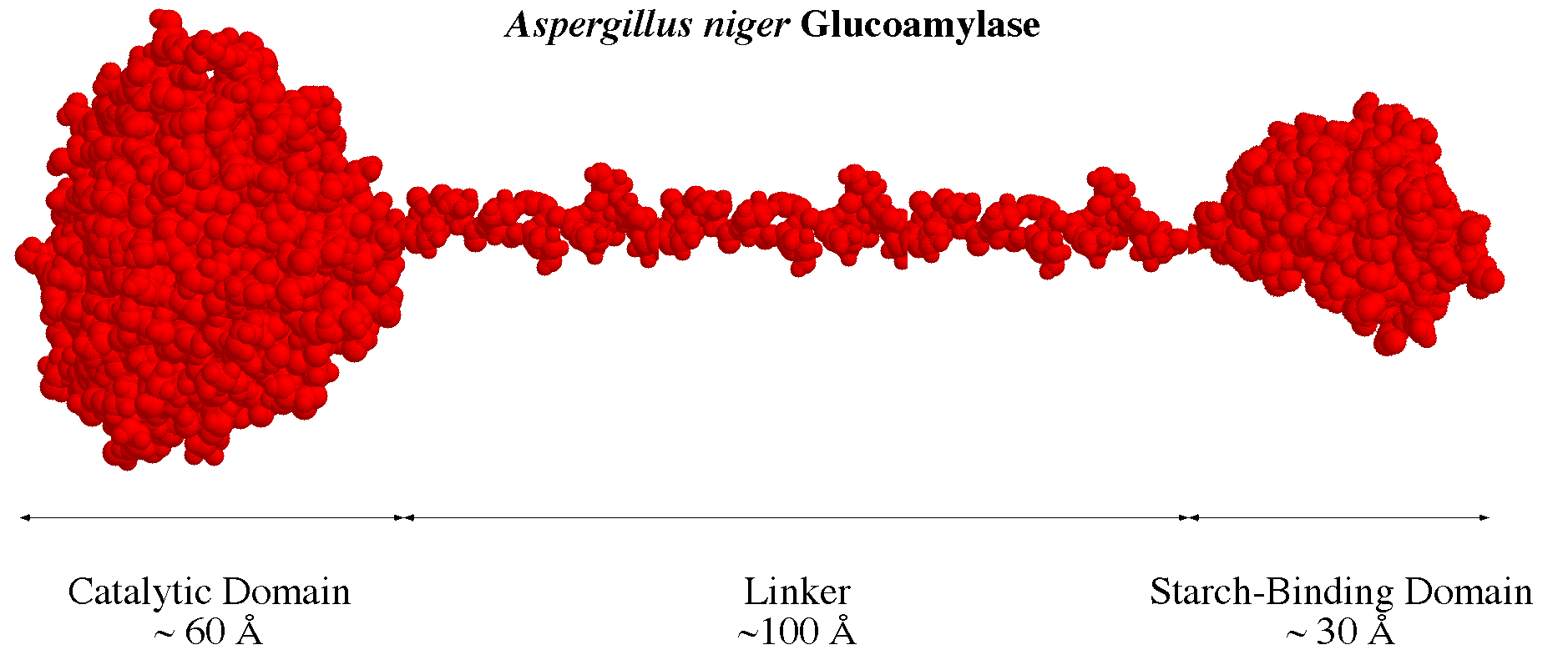
GA hydrolyzes 1,4-linked [[alpha]]-D-glucosyl residues from the nonreducing ends of oligo- and polysaccharide chains and can also hydrolyze [[alpha]]-1,6-D-glucosidic bonds. The ability of Aspergillus niger GA to catalyze these reactions is effectively used by the starch processing industry to produce glucose, an intermediate in HFCS production. Interest in improving some of the enzyme's process characteristics leads to a more intensive study of its structural and kinetic characteristics. The 3-D model of the CD of GA from Aspergillus awamori var. X100 (1gly) is an ([[alpha]]/[[alpha]])6 barrel surrounded by an extended O-glycosylated coil. Five sub-families of closely related GA sequences have been identified (Coutinho and Reilly, 1994a). The active site corresponds to five conserved sequence segments (S1-S5). The SBDs of fungal GAs are homologous to the C-terminal domain of Bacillus circulans no. 8 CGTase (3-D model 1cgt) (Klein and Schulz, 1991). A composite of Aspergillus niger GA based on these two models is given in Fig. 1.
Fig. 1. Possible structure of Aspergillus niger glucoamylase. The catalytic domain used was from 1gly, the linker was built from repetitions of the O-glycosylated belt around 1gly, and the starch-binding domain was domain E of 1cgt.
Hydrophobic cluster analysis (HCA), an efficient technique for the analysis and comparison of protein sequences when a 3-D structure is available, was applied to the study of the GA enzyme family (Coutinho and Reilly, 1994b). This resulted in an structure-based multisequence alignment. Studies of single and regional amino acid variations by comparing the 3-D model of proteins with protein sequence alignments is of major importance in understanding protein evolution and conducting protein engineering (Bordo, 1993).
We would like to increase of GA thermostability and selectivity and to change its optimal pH. A better understanding of GA characteristics provided by this study will support protein engineering of GA.
RESULTS AND DISCUSSION
Structure-based multisequence alignment - The alignment resulting from HCA study is shown in Fig. 2. The different sub-families show slight differences as observed before (Coutinho and Reilly, 1994b).
Fig. 2. HCA alignments visualized with Alscript (Barton, 1993). Secondary structures of 1gly and 1cgt as determined by Aleshin et al., 1992 (Ref. 1), Klein and Schulz, 1991 (Ref. 2), and for all other sequences as determined by HCA from primary sequences (Coutinho and Reilly, 1994b). For the SBD analysis, a consensus between strarch degrading enzymes and estimated secondary structure Svensson et al., 1989 (Ref 3.) and sites one (1) and two (2) for maltose binding are indicated. Black bars: [[alpha]]-helices; dark gray bars: 310-helices; light gray bars: [[beta]]-strands; boxed asterisks: turns. Large boxes: conserved regions (S1 to S5); vertical boxes: residues involved in interactions with substrate residues. Hydrophobic microdomain evaluation: italicized bold residues. Other relevant structural features: boxed C: Cys; boxed italicized N, S, or T: potentially glycosylated Asn, Ser, or Thr; dark boxes: confirmed disulfide bridges or glycosylated residues.
Fig. 3. Variability of amino acid residues in the catalytic domain of the Aspergillus glucoamylase sub-family using 1gly as reference: a) active site with the inhibitor acarbose (Aleshin et al., 1994); b) region opposite the active site. Scale: dark blue (no variability) to red (high variability).
Fig. 4. Variability of amino acid residues in the starch-binding domain of the Aspergillus glucoamylase sub-family using as reference 1cgt and maltose as bound to an equivalent CGTase (Lawson et al., 1994): a) site 1; b) site 2. Scale: dark blue (no variability) to red (high variability).
The active site region in the Aspergillus sub-family is very conserved, especially when compared with other regions of the CD. Some sites with high amino acid variability, eventually associated with differences in behavior of different GAs, can be found close to the active site. These sites are of major interest for protein engineering.
The aminoacid variability in the SBD of the different GAs, as depicted in the representation of the C-terminal domain of a CGTase, shows significant differences in site 1 and less amino acid variability in site 2. No other regions of the SBD appear to be conserved, an indication that the two major binding regions were found. The SBD seems less conserved through evolution, compared to he CD an indication of the SBD's lesser role in GA action.
ACKNOWLEDGMENTS
P.M.C. was supported by grant B.D./1057/90-IF of Programa CIÊNCIA, Junta Nacional de Investigação Científica e Tecnológica, Lisboa, Portugal. We thank Drs. Alexander Aleshin and Richard Honzatko for providing structures of a refined 1gly and of the GA complex with acarbose.
REFERENCES
Aleshin, A., Golubev, A., Firsov, L. M. and Honzatko, R. B. (1992) J. Biol. Chem., 267, 19291-19298.
Aleshin, A., Firsov, L. M. and Honzatko, R. B. (1994) J. Biol. Chem., in press.
Barton, G. J. (1993) Protein Eng., 6, 37-40.
Bordo, D. (1993) CABIOS, 9, 639-645.
Coutinho, P. M. and Reilly, P. J. (1994a) Protein Eng., 7, 393-400.
Coutinho, P. M. and Reilly, P. J. (1994b) Protein Eng., in press.
Klein, C. and Schulz, G. E. (1991) J. Mol. Biol., 217, 737-750.
Lawson, C.L., Vanmontfort, R., Strokopytov, B., Rozeboom, H.J., Kalk, K.H., Devries, G.E., Penninga, D., Dijkhuizen, L., and Dijkstra, B.W. (1994), J. Mol. Biol., 236, 590-600.
Svensson, B., Jespersen, H., Sierks, M.R., and MacGregor, E.A. (1989), Biochem. J., 264, 309-311.
Last Changed: 06/23/94