Homework 5
Crystal structure of the Aequorea victoria green fluorescent protein
Ormoe, M; Cubitt, AB; Kallio, K; Gross, LA; Tsien, RY; Remington, SJ
SCIENCE (WASH.), vol. 273, no. 5280, pp. 1392-1395, 1996
3DB file
Abstract
Information
Rasmol Picture
Blue Emission
Abstract
The green fluorescent protein (GFP) from the Pacific Northwest jellyfish Aequorea victoria has generated intense interest as a marker for gene expression and localization of gene products. The chromophore, resulting from the spontaneous cyclization and oxidation of the sequence -Ser super(65) (or Thr super(65))-Tyr super(66)-Gly super(67)-, requires the native protein hold for both formation and fluorescence emission. The structure of Thr super(65) GFP has been determined at 1.9 angstrom resolution. The protein fold consists of an 11-stranded beta barrel with a coaxial helix, with the chromophore forming from the central helix. Directed mutagenesis of one residue adjacent to the chromophore, Thr super(203), to Tyr or His results in significantly red-shifted excitation and emission maxima.
Information
PDB ID number: 1EMA
Expression: Plasmid: Prsetb (Invitrogen) Expression_system: Escherichia Coli Expression_system_strain: Jm109 (De3)
Residue number: 236
Number of b-sheet: 11
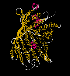
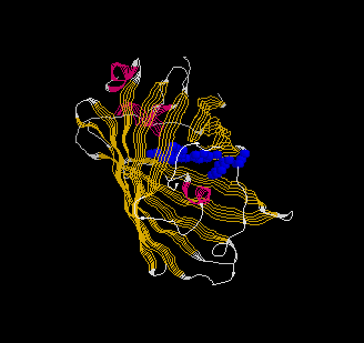
Rasmol Pictures
Overview
Chromophore(Ser65-His66-Gly67)
Blue Emission
Blue Emission Variant(Y66H/Y145F):homework3
Picture
Number of helix: 4
starting residue
ending residue
GLY 4
PHE 8
TRP 57
LEU 60
GLN 69
PHE 71